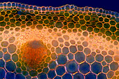
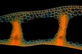
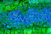
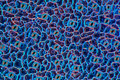
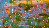
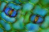
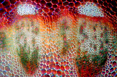
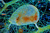
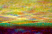
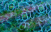
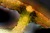
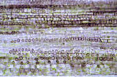
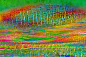
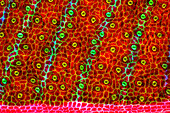
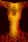
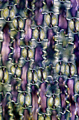
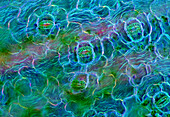
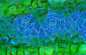
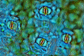
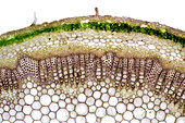
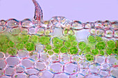
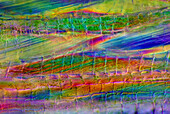
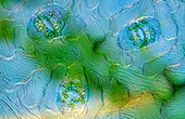
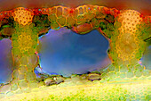
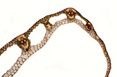
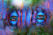
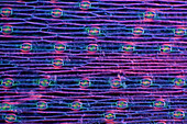
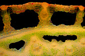
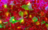
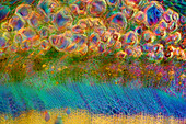
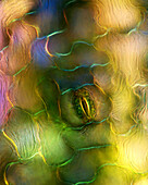
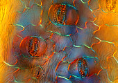
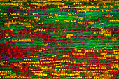
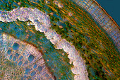
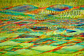
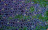
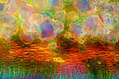
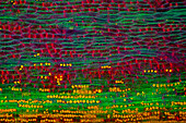
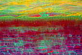
13925580 - The image presents Anemone sylvestris stalk in transversal cross-section, photographed through the microscope in polarized light at a magnification of 200X\n13925525 - The image presents Carex sp. leaf in transversal cross-section, photographed through the microscope in polarized light at a magnification of 100X\n13925473 - The image presents stomata in Spathiphyllum leaf epidermis, photographed through the microscope in polarized light at a magnification of 100X\n13925332 - The image presents stomata in hosta leaf epidermis, photographed through the microscope in polarized light at a magnification of 100X\n13925207 - The image presents stomata in Spathiphyllum leaf epidermis, photographed through the microscope in polarized light at a magnification of 400X\n13925148 - The image presents stomata in Spathiphyllum leaf epidermis, photographed through the microscope in polarized light at a magnification of 400X\n13925085 - The image presents vascular bundles in senecio stalk, photographed through the microscope in polarized light at a magnification of 200X\n13925083 - The image presents Utricularia trap, a kind of carnivorous plant, photographed through the microscope in polarized light and dark field, at a magnification of 100X\n13925010 - The image presents read leaf in transversal cross-section, photographed through the microscope in polarized light at a magnification of 100X\n13924337 - The image presents tissues in nettle stalk in longitudinal cross section, photographed through the microscope in polarized light at a magnification of 100X\n13925579 - The image presents stomata in Spathiphyllum leaf epidermis, photographed through the microscope in polarized light at a magnification of 200X\n13925392 - The image presents reed stalk in transversal cross-section, photographed through the microscope in polarized light at a magnification of 200X\n13925287 - The image presents tissues in nettle stalk in longitudinal cross-section, photographed through the microscope in polarized light at a magnification of 100X\n13925265 - The image presents nettle tissues in the stalk in longitudinal cross-section, photographed through the microscope in polarized light at a magnification of 100X\n13925100 - The image presents stomata in Stromanthe sp. leaf epidermis, photographed through the microscope in polarized light at a magnification of 100X\n13925093 - The image presents a single vascular bundle in Carex sp. stalk, photographed through the microscope in polarized light and dark field at a magnification of 200X\n13925078 - The image presents stomata in lily leaf epidermis, photographed through the microscope in polarized light at a magnification of 200X\n13925070 - The image presents stomata in Spathiphyllum sp. leaf epidermis, photographed through the microscope in polarized light at a magnification of 100X\n13925038 - The image presents stomata in Spathiphyllum leaf epidermis, photographed through the microscope in polarized light at a magnification of 200X\n13924900 - The image presents stomata in Spathiphyllum sp. leaf epidermis, photographed through the microscope in polarized light at a magnification of 100X\n13924866 - The image presents reed stalk in transversal cross-section, photographed through the microscope in polarized light at a magnification of 200X\n13924786 - The image presents knautia arvensis tissues in the transversal section of the stalk, photographed through the microscope in bright field, at a magnification of 100X\n13924748 - The image presents a single stoma in Knautia arvensis epidermis, photographed through the microscope in polarized light at a magnification of 200X\n13924555 - The image presents tissues in nettle stalk in longitudinal cross-section, photographed through the microscope in polarized light at a magnification of 100X\n13924552 - The image presents stomata in Spathiphyllum leaf epidermis, photographed through the microscope in polarized light at a magnification of 400X\n13924452 - The image presents reed stalk in transversal cross-section, photographed through the microscope in polarized light at a magnification of 200X\n13924434 - The image presents carex sp. leaf in transversal cross-section, photographed through the microscope in polarized light at a magnification of 100X\n13925468 - The image presents palisade mesophyll in hyacinthus leaf (transversal cross-section) photographed through the microscope in polarized light at a magnification of 200X\n13925303 - The image presents stomata in Spathiphyllum leaf epidermis, photographed through the microscope in polarized light at a magnification of 400X\n13925259 - The image presents stomata in hyacinth leaf epidermis, photographed through the microscope in polarized light at a magnification of 100X\n13925106 - The image presents reed stalk in the transversal cross-section, photographed through the microscope in polarized light and dark field, at a magnification of 100X\n13924924 - The image presents stomata in Croton leaf epidermis, photographed through the microscope in polarized light at a magnification of 200X\n13924809 - The image presents nettle tissues in the transversal cross-section of the stalk, photographed through the microscope in polarized light at a magnification of 200X\n13924732 - The image presents a single stoma in Spathiphyllum leaf epidermis, photographed through the microscope in polarized light at a magnification of 200X\n13924539 - The image presents stomata in Spathiphyllum leaf epidermis, photographed through the microscope in polarized light at a magnification of 200X\n13924490 - The image presents tissues in nettle stalk in longitudinal cross-section, photographed through the microscope in polarized light at a magnification of 100X. The round yellow structures are druses. Druses are the structures created by calcium oxalate.\n13925603 - The image presents oak tissues in transversal cross-section of the stalk, photographed through the microscope in polarized light at a magnification of 100X\n13925454 - The image presents nettle tissues in the stalk in longitudinal cross-section, photographed through the microscope at a magnification of 100X\n13925048 - The image presents stomata in Croton leaf epidermis, photographed through the microscope in polarized light at a magnification of 100X\n13925009 - The image presents nettle stalk longitudinal cross-section photographed through the microscope in polarized light at a magnification of 100X\n13924796 - The image presents knautia arvensis tissues in the transversal section of the stalk, photographed through the microscope in bright field, at a magnification of 100X\n13924698 - The image presents nettle tissues in the stalk in longitudinal cross-section, photographed through the microscope at a magnification of 100X\n13925464 - The image presents stomata in Stromanthe sp. leaf epidermis, photographed through the microscope in polarized light at a magnification of 100X\n13925405 - The image presents stomata in Spathiphyllum sp. leaf epidermis, photographed through the microscope in polarized light at a magnification of 100X\n13925240 - The image presents stomata in Spathiphyllum leaf epidermis, photographed through the microscope in polarized light at a magnification of 200X\n13925092 - The image presents oak xylem tissue in the transversal cross-section of the stalk, photographed through the microscope in polarized light at a magnification of 400X\n13924921 - The image presents senecio tissues in the transversal cross-section through the stalk, photographed through the microscope in polarized light and dark field at a magnification of 100X\n13924913 - The image presents stomata in Spathiphyllum leaf epidermis, photographed through the microscope in polarized light at a magnification of 200X\n13924624 - The image presents a single stoma in Spathiphyllum sp. leaf epidermis, photographed through the microscope in polarized light at a magnification of 200X\n13924564 - The image presents stomata in Spathiphyllum leaf epidermis, photographed through the microscope in polarized light at a magnification of 200X\n13924392 - Tissues in nettle stalk photographed through the microscope\n13924302 - The image presents nettle tissues in the transversal section of the stalk, photographed through the microscope in polarized light at a magnification of 400X\n13925616 - The image presents stomata in Stromanthe sp. leaf epidermis, photographed through the microscope in polarized light at a magnification of 100X\n13925549 - The image presents a small part of forsythia stalk tissues in transversal cross-section, photographed through the microscope in polarized light at a magnification of 100X\n13925345 - The image presents nettle tissues in the stalk in transversall cross-section, photographed through the microscope in polarized light and dark field at a magnification of 100X.\n13925297 - The image presents stomata in Spathiphyllum leaf epidermis, photographed through the microscope in polarized light at a magnification of 100X\n13925241 - The image presents nettle tisues in the transversal cross-section of the stalk, photographed through the microscope in polarized light at a magnification of 200X\n13924841 - The image presents tissues in nettle stalk in longitudinal cross-section, photographed through the microscope in polarized light at a magnification of 100X. The round yellow structures on the bottom are druses. Druses are the structures created by calcium oxalat.\n13924686 - The image presents nettle tissues in longitudinal cross-section of the stalk, photographed through the microscope in polarized light at a magnification of 100X. Small, round orange particles are the crystals of calcium oxalate called druses.\n13924655 - The image presents stomata in lily leaf epidermis, photographed through the microscope in polarized light at a magnification of 200X\n13924580 - The image presents reed stalk in transversal cross-section, photographed through the microscope in bright field, at a magnification of 100X\n70217764 - Allergic woman70293703 - Travelling by boat from Dubrovnik to Mljet island, Mljet island is in background, Croatia70103060 - yes, Cold, Colds, Color, Colour, Dark-haired, Disease, Diseases, Female, Generation X, Handkerchief71335930 - Itsukushima Miyajima Japan. Handkerchiefs70217763 - Allergic woman