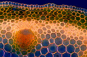
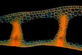
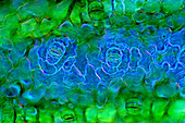
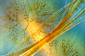
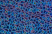
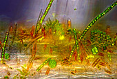
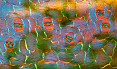
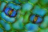
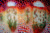
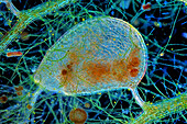
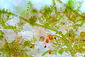
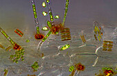
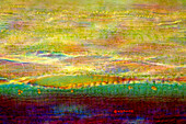
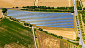
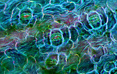
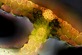
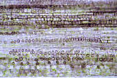
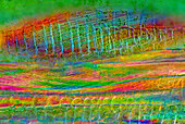
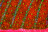
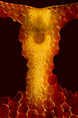
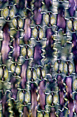
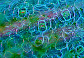
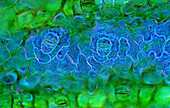
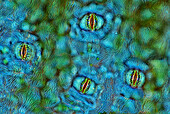
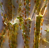
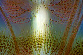
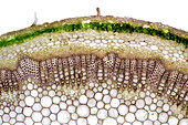
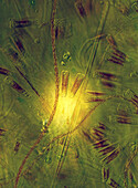
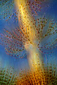
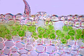
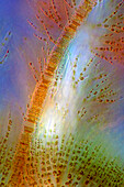
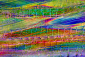
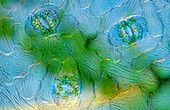
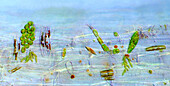
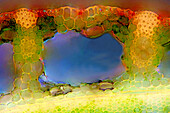
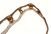
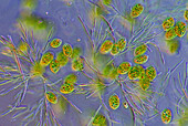
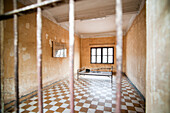
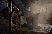
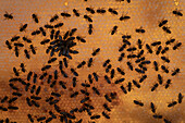
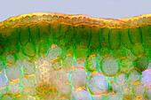
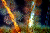
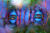
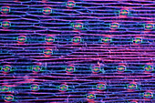
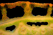
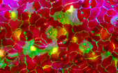
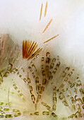
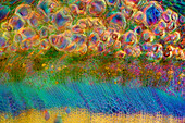
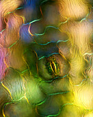
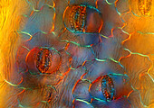
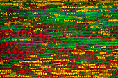
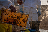
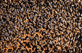
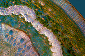
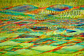
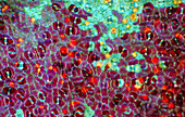
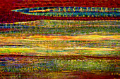
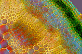
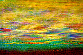
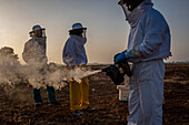
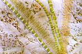
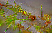
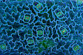
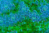
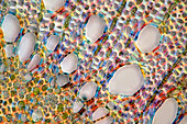
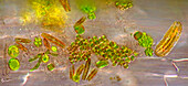
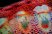
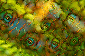
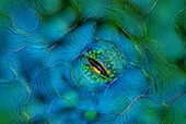
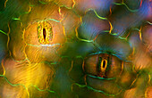
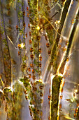
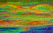
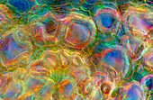
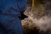
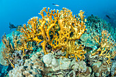
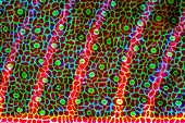
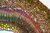
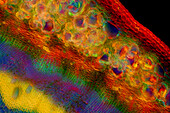
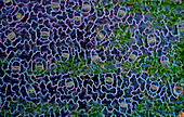
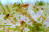
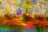
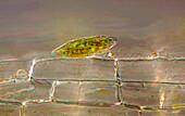
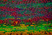
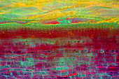
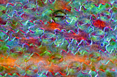
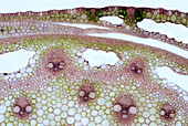
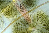
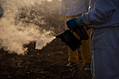
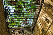
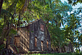
71441015 - A ground mounted system of many solar panels on an open space with trees and clouds in the background, Hesse, Germany13925580 - The image presents Anemone sylvestris stalk in transversal cross-section, photographed through the microscope in polarized light at a magnification of 200X\n13925525 - The image presents Carex sp. leaf in transversal cross-section, photographed through the microscope in polarized light at a magnification of 100X\n13925473 - The image presents stomata in Spathiphyllum leaf epidermis, photographed through the microscope in polarized light at a magnification of 100X\n13925385 - The image presents Fragilaria sp., a kind of diatoms against Batrachospermum, a kind of red algae, photographed through the microscope in polarized light at a magnification of 200X\n13925332 - The image presents stomata in hosta leaf epidermis, photographed through the microscope in polarized light at a magnification of 100X\n13925278 - The image presents various tiny algae settled on Lemna sp. root, photographed through the microscope in polarized light at a magnification of 400X. On the right are visible diatoms closed in a special protecting case.\n13925207 - The image presents stomata in Spathiphyllum leaf epidermis, photographed through the microscope in polarized light at a magnification of 400X\n13925148 - The image presents stomata in Spathiphyllum leaf epidermis, photographed through the microscope in polarized light at a magnification of 400X\n13925085 - The image presents vascular bundles in senecio stalk, photographed through the microscope in polarized light at a magnification of 200X\n13925083 - The image presents Utricularia trap, a kind of carnivorous plant, photographed through the microscope in polarized light and dark field, at a magnification of 100X\n13925044 - The image presents various tiny algae settled on Lemna sp. root, photographed through the microscope in polarized light at a magnification of 200X\n13925041 - The image presents various tiny algae settled on Lemna sp. root, photographed through the microscope in polarized light at a magnification of 200X\n13925010 - The image presents read leaf in transversal cross-section, photographed through the microscope in polarized light at a magnification of 100X\n13924337 - The image presents tissues in nettle stalk in longitudinal cross section, photographed through the microscope in polarized light at a magnification of 100X\n13826937 - beekeeping , apiculture, Carmona, Andalucia, Spain71441014 - Aerial view of a ground-mounted system with photovoltaic modules next to a road and railway line, Hesse, Germany13925579 - The image presents stomata in Spathiphyllum leaf epidermis, photographed through the microscope in polarized light at a magnification of 200X\n13925392 - The image presents reed stalk in transversal cross-section, photographed through the microscope in polarized light at a magnification of 200X\n13925287 - The image presents tissues in nettle stalk in longitudinal cross-section, photographed through the microscope in polarized light at a magnification of 100X\n13925265 - The image presents nettle tissues in the stalk in longitudinal cross-section, photographed through the microscope in polarized light at a magnification of 100X\n13925100 - The image presents stomata in Stromanthe sp. leaf epidermis, photographed through the microscope in polarized light at a magnification of 100X\n13925093 - The image presents a single vascular bundle in Carex sp. stalk, photographed through the microscope in polarized light and dark field at a magnification of 200X\n13925078 - The image presents stomata in lily leaf epidermis, photographed through the microscope in polarized light at a magnification of 200X\n13925070 - The image presents stomata in Spathiphyllum sp. leaf epidermis, photographed through the microscope in polarized light at a magnification of 100X\n13925038 - The image presents stomata in Spathiphyllum leaf epidermis, photographed through the microscope in polarized light at a magnification of 200X\n13924900 - The image presents stomata in Spathiphyllum sp. leaf epidermis, photographed through the microscope in polarized light at a magnification of 100X\n13924866 - The image presents reed stalk in transversal cross-section, photographed through the microscope in polarized light at a magnification of 200X\n13924859 - "The image presents Cladophora sp. ""twigs"" (a kind of green algae) with Cocconeis sp. (a kin of diatoms) settled on it, photographed through the microscope in polarized light at a magnification of 200X"\n13924803 - The image presents Fragilaria sp., a kind of diatoms against Batrachospermum, a kind of red algae, photographed through the microscope in polarized light at a magnification of 200X\n13924786 - The image presents knautia arvensis tissues in the transversal section of the stalk, photographed through the microscope in bright field, at a magnification of 100X\n13924785 - The image presents diatoms (mostly Gomphonema sp.) photographed through the microscope in polarized light at a magnification of 200X\n13924764 - The image presents Batrachospermum sp., a kind of red algae, photographed through the microscope in polarized light at a magnification of 200X\n13924748 - The image presents a single stoma in Knautia arvensis epidermis, photographed through the microscope in polarized light at a magnification of 200X\n13924602 - The image presents Fragilaria sp., a kind of diatoms and Batrachospermum sp., a kind of red algae, photographed through the microscope in polarized light at a magnification of 200X\n13924555 - The image presents tissues in nettle stalk in longitudinal cross-section, photographed through the microscope in polarized light at a magnification of 100X\n13924552 - The image presents stomata in Spathiphyllum leaf epidermis, photographed through the microscope in polarized light at a magnification of 400X\n13924513 - The image presents various tiny algae settled on Lemna sp. root, photographed through the microscope in polarized light at a magnification of 200X\n13924452 - The image presents reed stalk in transversal cross-section, photographed through the microscope in polarized light at a magnification of 200X\n13924434 - The image presents carex sp. leaf in transversal cross-section, photographed through the microscope in polarized light at a magnification of 100X\n13924404 - The image presents euglenoids among green algae, photographed through the microscope in polarized light at a magnification of 100X\n13848559 - Torture Room at The Tuol Sleng Genocide Museum (S-21 Prison), Phnom Penh, Cambodia. The Tuol Sleng Genocide Museum (S-21 Prison) is a school that was converted into a prison during the rule of the Khmer Rouge and is an extremely interesting way to learn about Cambodias shocking history.13826936 - beekeeping , apiculture, Carmona, Andalucia, Spain13826935 - beekeeping , apiculture, Carmona, Andalucia, Spain13925468 - The image presents palisade mesophyll in hyacinthus leaf (transversal cross-section) photographed through the microscope in polarized light at a magnification of 200X\n13925399 - The image presents Batrachospermum sp., a kind of red algae, and FRagilaria sp., a kind of diatoms, photographed through the microscope in polarized light and dark field at a magnification of 100X\n13925303 - The image presents stomata in Spathiphyllum leaf epidermis, photographed through the microscope in polarized light at a magnification of 400X\n13925259 - The image presents stomata in hyacinth leaf epidermis, photographed through the microscope in polarized light at a magnification of 100X\n13925106 - The image presents reed stalk in the transversal cross-section, photographed through the microscope in polarized light and dark field, at a magnification of 100X\n13924924 - The image presents stomata in Croton leaf epidermis, photographed through the microscope in polarized light at a magnification of 200X\n13924902 - The image presents Batrachospermum sp., a kind of red algae and some kind of diatoms photographed through the microscope in slightly polarized light at a magnification of 200X\n13924809 - The image presents nettle tissues in the transversal cross-section of the stalk, photographed through the microscope in polarized light at a magnification of 200X\n13924732 - The image presents a single stoma in Spathiphyllum leaf epidermis, photographed through the microscope in polarized light at a magnification of 200X\n13924539 - The image presents stomata in Spathiphyllum leaf epidermis, photographed through the microscope in polarized light at a magnification of 200X\n13924490 - The image presents tissues in nettle stalk in longitudinal cross-section, photographed through the microscope in polarized light at a magnification of 100X. The round yellow structures are druses. Druses are the structures created by calcium oxalate.\n13826934 - beekeeping , apiculture, Carmona, Andalucia, Spain13826933 - beekeeping , apiculture, Carmona, Andalucia, Spain13925603 - The image presents oak tissues in transversal cross-section of the stalk, photographed through the microscope in polarized light at a magnification of 100X\n13925454 - The image presents nettle tissues in the stalk in longitudinal cross-section, photographed through the microscope at a magnification of 100X\n13925048 - The image presents stomata in Croton leaf epidermis, photographed through the microscope in polarized light at a magnification of 100X\n13925009 - The image presents nettle stalk longitudinal cross-section photographed through the microscope in polarized light at a magnification of 100X\n13924796 - The image presents knautia arvensis tissues in the transversal section of the stalk, photographed through the microscope in bright field, at a magnification of 100X\n13924698 - The image presents nettle tissues in the stalk in longitudinal cross-section, photographed through the microscope at a magnification of 100X\n13826932 - beekeeping , apiculture, Carmona, Andalucia, Spain13925530 - The image presents various algae (Fragilaria sp., a kind of diatoms, Batrachospermum sp., a kind of red algae and filamentous green algae) photographed through the microscope in slightly polarized light at a magnification of 200X\n13925464 - The image presents stomata in Stromanthe sp. leaf epidermis, photographed through the microscope in polarized light at a magnification of 100X\n13925443 - The image presents various tiny algae settled on Lemna sp. root, photographed through the microscope in polarized light at a magnification of 200X\n13925405 - The image presents stomata in Spathiphyllum sp. leaf epidermis, photographed through the microscope in polarized light at a magnification of 100X\n13925240 - The image presents stomata in Spathiphyllum leaf epidermis, photographed through the microscope in polarized light at a magnification of 200X\n13925092 - The image presents oak xylem tissue in the transversal cross-section of the stalk, photographed through the microscope in polarized light at a magnification of 400X\n13924948 - The image presents various tiny algae settled on Lemna sp. root, photographed through the microscope in polarized light at a magnification of 400X. On the right are visible diatoms closed in a special protecting case.\n13924921 - The image presents senecio tissues in the transversal cross-section through the stalk, photographed through the microscope in polarized light and dark field at a magnification of 100X\n13924913 - The image presents stomata in Spathiphyllum leaf epidermis, photographed through the microscope in polarized light at a magnification of 200X\n13924624 - The image presents a single stoma in Spathiphyllum sp. leaf epidermis, photographed through the microscope in polarized light at a magnification of 200X\n13924564 - The image presents stomata in Spathiphyllum leaf epidermis, photographed through the microscope in polarized light at a magnification of 200X\n13924504 - "The image presents Cladophora sp. ""twigs"" (a kind of green algae) with Cocconeis sp. (a kin of diatoms) settled on it, photographed through the microscope in polarized light at a magnification of 100X"\n13924392 - Tissues in nettle stalk photographed through the microscope\n13924302 - The image presents nettle tissues in the transversal section of the stalk, photographed through the microscope in polarized light at a magnification of 400X\n13826930 - beekeeping , apiculture, Carmona, Andalucia, Spain13990877 - Fire coral (Millepora dichotoma) are similar to coral but are not true corals. They are instead more closely related to hydrozoans,making them hydrocorals,Yap,Federated States of Micronesia13925616 - The image presents stomata in Stromanthe sp. leaf epidermis, photographed through the microscope in polarized light at a magnification of 100X\n13925549 - The image presents a small part of forsythia stalk tissues in transversal cross-section, photographed through the microscope in polarized light at a magnification of 100X\n13925345 - The image presents nettle tissues in the stalk in transversall cross-section, photographed through the microscope in polarized light and dark field at a magnification of 100X.\n13925297 - The image presents stomata in Spathiphyllum leaf epidermis, photographed through the microscope in polarized light at a magnification of 100X\n13925243 - The image presents various tiny algae settled on Lemna sp. root, photographed through the microscope in polarized light at a magnification of 200X\n13925241 - The image presents nettle tisues in the transversal cross-section of the stalk, photographed through the microscope in polarized light at a magnification of 200X\n13924997 - The image presents diatoms closed in a special kind of the protecting case, photographed through the microscope in polarized light at a magnification of 400X\n13924841 - The image presents tissues in nettle stalk in longitudinal cross-section, photographed through the microscope in polarized light at a magnification of 100X. The round yellow structures on the bottom are druses. Druses are the structures created by calcium oxalat.\n13924686 - The image presents nettle tissues in longitudinal cross-section of the stalk, photographed through the microscope in polarized light at a magnification of 100X. Small, round orange particles are the crystals of calcium oxalate called druses.\n13924655 - The image presents stomata in lily leaf epidermis, photographed through the microscope in polarized light at a magnification of 200X\n13924580 - The image presents reed stalk in transversal cross-section, photographed through the microscope in bright field, at a magnification of 100X\n13924544 - The image presents Batrachospermum sp., a kind of red algae and Fragilaria sp., a kind of diatoms, photographed through the microscope in polarized light at a magnification of 200X\n13826931 - beekeeping , apiculture, Carmona, Andalucia, Spain13885395 - Scenic view of a woman taking a picture with her smart phone, standing on a cliff-side slope in front of a series of waterfalls flowing from the craggy cliffs of the East Fjords, making her appear small against the vast landscape in front of her; East Iceland, Iceland14306175 - Ruined cells of penal colony on Ile Saint-Joseph, Iles du Salut (Islands of Salvation), French Guiana, Overseas department and region of France, French Guiana, South America14306173 - Ruined cells of penal colony on Ile Saint-Joseph, Iles du Salut (Islands of Salvation), French Guiana, Overseas department and region of France, French Guiana, South America70227069 - solar panels on the grass roof of the Willy-Brandt Haus headquarters of the SPD Social Democratic party Berlin, Germany70105287 - 16 year old girl in phone booth, Porto. Portugal70131531 - English telephone booth at Whyte Avenue, Old Strathcona area. Edmonton. Alberta, Canadapage suivante