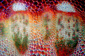
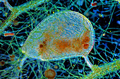
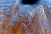
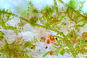
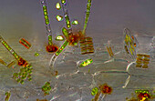
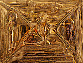
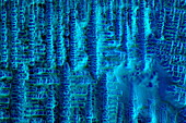
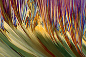
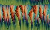
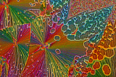
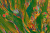
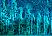
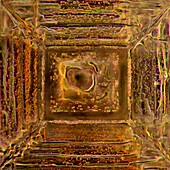
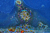
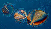
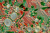
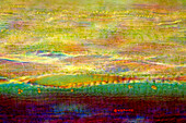
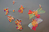
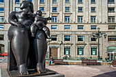
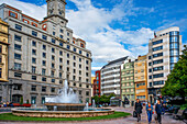
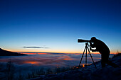
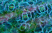
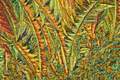
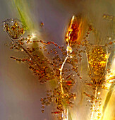
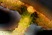
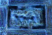
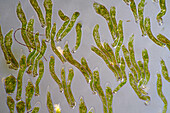
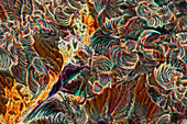
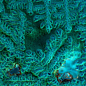
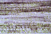
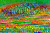
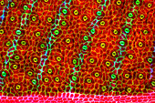
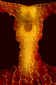
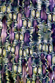
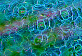
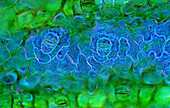
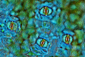
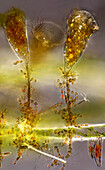
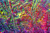
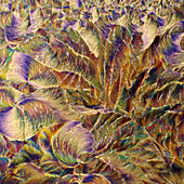
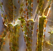
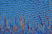
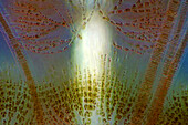
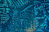
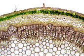
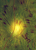
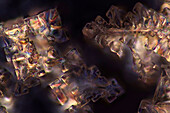
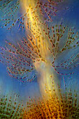
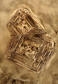
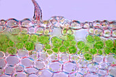
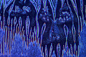
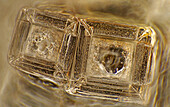
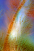
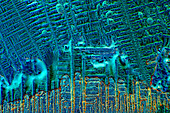
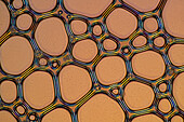
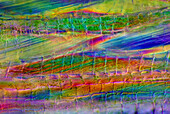
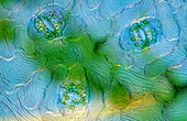
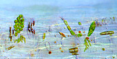
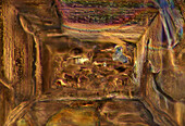
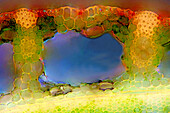
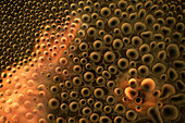
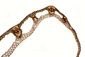
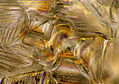
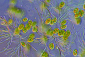
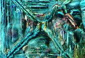
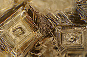
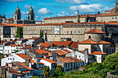
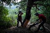
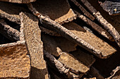
13925085 - The image presents vascular bundles in senecio stalk, photographed through the microscope in polarized light at a magnification of 200X\n13925083 - The image presents Utricularia trap, a kind of carnivorous plant, photographed through the microscope in polarized light and dark field, at a magnification of 100X\n13925079 - The image presents crystallized mixture of kitchen salt and erythritol, photographed through the microscope in polarized light at a magnification of 100X\n13925044 - The image presents various tiny algae settled on Lemna sp. root, photographed through the microscope in polarized light at a magnification of 200X\n13925041 - The image presents various tiny algae settled on Lemna sp. root, photographed through the microscope in polarized light at a magnification of 200X\n13925035 - The image presents crystallized mixture of kitchen salt and erythritol, photographed through the microscope in polarized light at a magnification of 100X\n13925027 - The image presents crystallized soy sauce, photographed through the microscope in polarized light at a magnification of 100X\n13925010 - The image presents read leaf in transversal cross-section, photographed through the microscope in polarized light at a magnification of 100X\n13924998 - The image presents crystallized resorcinol, photographed through the microscope in polarized light at a magnification of 100X\n13924862 - The image presents a single crystal of recrystallized kitchen salt, photographed through the microscope in polarized light at a magnification of 200X\n13924844 - The image presents crystallized mixture of urea and paracetamol, photographed through the microscope in polarized light at a magnification of 100X\n13924827 - The image presents crystallized soy sauce, photographed through the microscope in polarized light at a magnification of 100X\n13924810 - The image presents crystallized soy sauce, photographed through the microscope in polarized light at a magnification of 100X\n13924754 - The image presents crystallized mixture of malic acid and hydroquinone photographed through the microscope in polarized light at a magnification of 100X\n13924619 - The image presents crystallized tartaric acid, photographed through the microscope in polarized light at a magnification of 100X\n13924584 - The image presents crystallized soy sauce, photographed through the microscope in polarized light at a magnification of 100X\n13924550 - The image presents a single crystal of recrystallized kitchen salt, photographed through the microscope in polarized light at a magnification of 200X\n13924465 - The image presents crystallized silver nitrate, photographed through the microscope in polarized light at a magnification of 100X\n13924437 - The image presents crystallized sulfur, photographed through the microscope in polarized light at a magnification of 100X\n13924395 - The image presents crystallized tartaric acid, photographed through the microscope in polarized light at a magnification of 100X\n13924337 - The image presents tissues in nettle stalk in longitudinal cross section, photographed through the microscope in polarized light at a magnification of 100X\n13924325 - The image presents crystallized mixture of malic acid, salicylic acid and acetanilid, photographed through the microscope in polarized light at a magnification of 100X\n13897585 - Portrait of a young beautiful caucasian woman in her 20´s with blue eyes photographing with an old vintage camera on a tripod outdoor. Lifestyle concept.13834173 - Organ inside Cathedral of Santiago de Compostela at Praza do Obradoiro Santiago de Compostela A Coruña, Spain.13834092 - Maternity, by Fernando Botero. Escandalera Square, Oviedo, Spain13834090 - Cajastur bank building and fountain on the Plaza de la Escandalera in Oviedo in Asturias region, Spain13827060 - Cork collecting Natural Park Los Alcornocales Cortes de la Frontera Andalusia Malaga Spain13827059 - Cork collecting Natural Park Los Alcornocales Cortes de la Frontera Andalusia Malaga Spain13827041 - Cork collecting Natural Park Los Alcornocales Cortes de la Frontera Andalusia Malaga Spain13827025 - Cork collecting Natural Park Los Alcornocales Cortes de la Frontera Andalusia Malaga Spain13826039 - Tourists, in Wat Arun (Temple of Dawn), also called Wat Bangmakok Noek, Bangkok, Thailand13825338 - Grutas de Sao Vicente.Madeira, Portugal13824575 - Taj Mahal and roofs of the city, Agra, India13824312 - Tourists watching sunrise at Angkor Wat, Siem Reap, Cambodia13824038 - Poporo Quimbaya, Pre-Columbian goldwork collection, Gold museum, Museo del Oro, Bogota, Colombia, America13824009 - Visitors. Right `Canasta de frutas´. Left `Mujer delante de una ventana´.Botero Museum, Bogota, Colombia13823546 - Old town, Salzburg, Austria13793521 - Composite Grizzly Stands In Front Of Lake With Mt. Mckinley In The Background, Alaska Composite13793438 - Silhouette Of Photographer At Sunset In Eagle River Valley, Southcentral, Alaska, Winter13925579 - The image presents stomata in Spathiphyllum leaf epidermis, photographed through the microscope in polarized light at a magnification of 200X\n13925520 - The image presents crystallized callus remover, photographed through the microscope in polarized light at a magnification of 100X.\n13925423 - The image presentstwo suctorians ( a kind of ciliate) and tiny diatoms, photographed through the microscope in polarized light at a magnification of 200X\n13925392 - The image presents reed stalk in transversal cross-section, photographed through the microscope in polarized light at a magnification of 200X\n13925377 - The image presents a single crystal of recrystallized salt, photographed through the microscope in polarized light at a magnification of 100X\n13925372 - The image presents a single crystal of recrystallized kitchen salt, photographed through the microscope in polarized light at a magnification of 200X\n13925367 - The image presents Ophrydium sp. ( a kind of colonial ciliates), photographed through the microscope in polarized light at a magnification of 100X\n13925342 - The image presents crystallized malic acid, photographed through the microscope in polarized light at a magnification of 100X\n13925338 - The image presents crystallized soy sauce, photographed through the microscope in polarized light at a magnification of 100X\n13925287 - The image presents tissues in nettle stalk in longitudinal cross-section, photographed through the microscope in polarized light at a magnification of 100X\n13925265 - The image presents nettle tissues in the stalk in longitudinal cross-section, photographed through the microscope in polarized light at a magnification of 100X\n13925174 - The image presents crystallized tartaric acid, photographed through the microscope in polarized light at a magnification of 100X\n13925104 - The image presents diptera eggs, photographed through the microscope in polarized light at a magnification of 100X\n13925100 - The image presents stomata in Stromanthe sp. leaf epidermis, photographed through the microscope in polarized light at a magnification of 100X\n13925093 - The image presents a single vascular bundle in Carex sp. stalk, photographed through the microscope in polarized light and dark field at a magnification of 200X\n13925078 - The image presents stomata in lily leaf epidermis, photographed through the microscope in polarized light at a magnification of 200X\n13925070 - The image presents stomata in Spathiphyllum sp. leaf epidermis, photographed through the microscope in polarized light at a magnification of 100X\n13925038 - The image presents stomata in Spathiphyllum leaf epidermis, photographed through the microscope in polarized light at a magnification of 200X\n13924900 - The image presents stomata in Spathiphyllum sp. leaf epidermis, photographed through the microscope in polarized light at a magnification of 100X\n13924894 - The image presentstwo suctorians ( a kind of ciliate) and tiny diatoms, photographed through the microscope in polarized light at a magnification of 200X\n13924892 - The image presents crystallized mixture of urea, paracetamol and resorcinol, photographed through the microscope in polarized light at a magnification of 100X\n13924873 - The image presents crystallized mixture of malic acid, salicylic acid and acetanilid, photographed through the microscope in polarized light at a magnification of 100X\n13924866 - The image presents reed stalk in transversal cross-section, photographed through the microscope in polarized light at a magnification of 200X\n13924859 - "The image presents Cladophora sp. ""twigs"" (a kind of green algae) with Cocconeis sp. (a kin of diatoms) settled on it, photographed through the microscope in polarized light at a magnification of 200X"\n13924829 - The image presents crystallized mixture of erythritol and TRIS, photographed through the microscope in polarized light at a magnification of 100X\n13924803 - The image presents Fragilaria sp., a kind of diatoms against Batrachospermum, a kind of red algae, photographed through the microscope in polarized light at a magnification of 200X\n13924794 - The image presents crystallized soy sauce, photographed through the microscope in polarized light at a magnification of 100X\n13924786 - The image presents knautia arvensis tissues in the transversal section of the stalk, photographed through the microscope in bright field, at a magnification of 100X\n13924785 - The image presents diatoms (mostly Gomphonema sp.) photographed through the microscope in polarized light at a magnification of 200X\n13924768 - The image presents crystallized mixture os sugar and salt, photographed through the microscope in polarized light at a magnification of 100X\n13924764 - The image presents Batrachospermum sp., a kind of red algae, photographed through the microscope in polarized light at a magnification of 200X\n13924752 - The image presents crystals of recrystallized kitchen salt, photographed through the microscope in polarized light at a magnification of 100X\n13924748 - The image presents a single stoma in Knautia arvensis epidermis, photographed through the microscope in polarized light at a magnification of 200X\n13924747 - The image presents crystallized soy sauce photographed through the microscope in polarized light and phase contrast at a magnification of 100X\n13924612 - The image presents crystals of recrystallized kitchen salt, photographed through the microscope in polarized light at a magnification of 100X\n13924607 - The image presents crystallized mixture of urea and resorcinol, photographed through the microscope in polarized light at a magnification of 100X\n13924602 - The image presents Fragilaria sp., a kind of diatoms and Batrachospermum sp., a kind of red algae, photographed through the microscope in polarized light at a magnification of 200X\n13924599 - The image presents crystallized soy sauce, photographed through the microscope in polarized light at a magnification of 100X\n13924573 - The image presents air bubbles formed in foemaed milk photographed through the microscope in polarized light at a magnification of 100X\n13924555 - The image presents tissues in nettle stalk in longitudinal cross-section, photographed through the microscope in polarized light at a magnification of 100X\n13924552 - The image presents stomata in Spathiphyllum leaf epidermis, photographed through the microscope in polarized light at a magnification of 400X\n13924513 - The image presents various tiny algae settled on Lemna sp. root, photographed through the microscope in polarized light at a magnification of 200X\n13924489 - The image presents crystallized resorcinol, photographed through the microscope in polarized light at a magnification of 100X\n13924487 - The image presents crystallized mixture of kitchen salt and erythritol, photographed through the microscope in polarized light at a magnification of 100X\n13924452 - The image presents reed stalk in transversal cross-section, photographed through the microscope in polarized light at a magnification of 200X\n13924442 - The image presents tiny air bubbles photographed through the microscope in polarized light at a magnification of 100X\n13924434 - The image presents carex sp. leaf in transversal cross-section, photographed through the microscope in polarized light at a magnification of 100X\n13924422 - The image presents crystallized mixture of kitchen salt and erythritol, photographed through the microscope in polarized light at a magnification of 100X\n13924404 - The image presents euglenoids among green algae, photographed through the microscope in polarized light at a magnification of 100X\n13924398 - The image presents recrystallized sugar photographed through the microscope in polarized light at a magnification of 100X\n13924389 - The image presents recrystallized mixture of salt and erithrytol, photographed through the microscope in polarized light at a magnification of 100X\n13924305 - The image presents crystals of recrystallized kitchen salt, photographed through the microscope in polarized light at a magnification of 100X\n13900408 - Park and National Palace of Pena (Palacio de la Pena), Sintra, Portugal13898836 - Portrait of a young beautiful caucasian woman in her 20´s with blue eyes photographing with an old vintage camera on a tripod outdoor. Lifestyle concept.13898745 - Portrait of a young beautiful caucasian woman in her 20´s with blue eyes photographing with an old vintage camera on a tripod outdoor. Lifestyle concept.13834186 - Old town and Cathedral of Santiago de Compostela at Praza do Obradoiro Santiago de Compostela A Coruña, Spain.13827057 - Cork collecting Natural Park Los Alcornocales Cortes de la Frontera Andalusia Malaga Spain13827040 - Cork collecting Natural Park Los Alcornocales Cortes de la Frontera Andalusia Malaga Spain13827023 - Cork collecting Natural Park Los Alcornocales Cortes de la Frontera Andalusia Malaga Spain13826759 - Chamberlain Square, Birmingham, Englandnext page